Gurtner Lab, 2019.
Microscopy
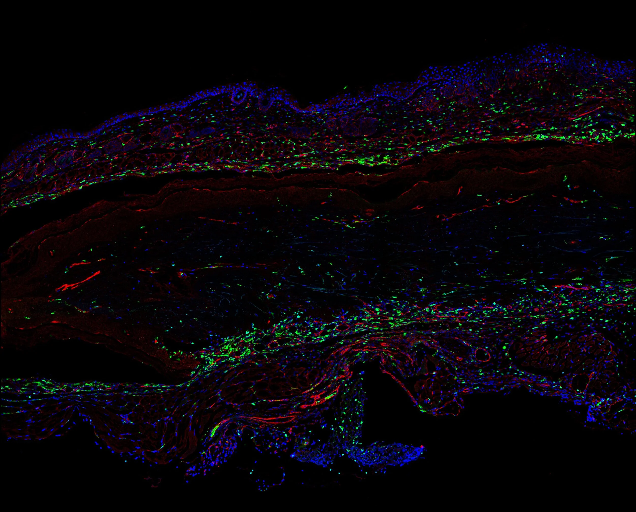
The image above shows in blue, mouse skin cells of different types (epidermis, dermis, and muscle fascia), and in red, the cross sections of blood vessels within the skin at 10X magnification. A confocal microscope is used to achieve high quality fluorescence images by using a pinhole technique: out of focus light is blocked out during image formation. Each took roughly 2 minutes to capture.
Each image provides a deeper understanding of the deeply rooted and naturally abundant convergences between scientific discovery and artistic aesthetics.
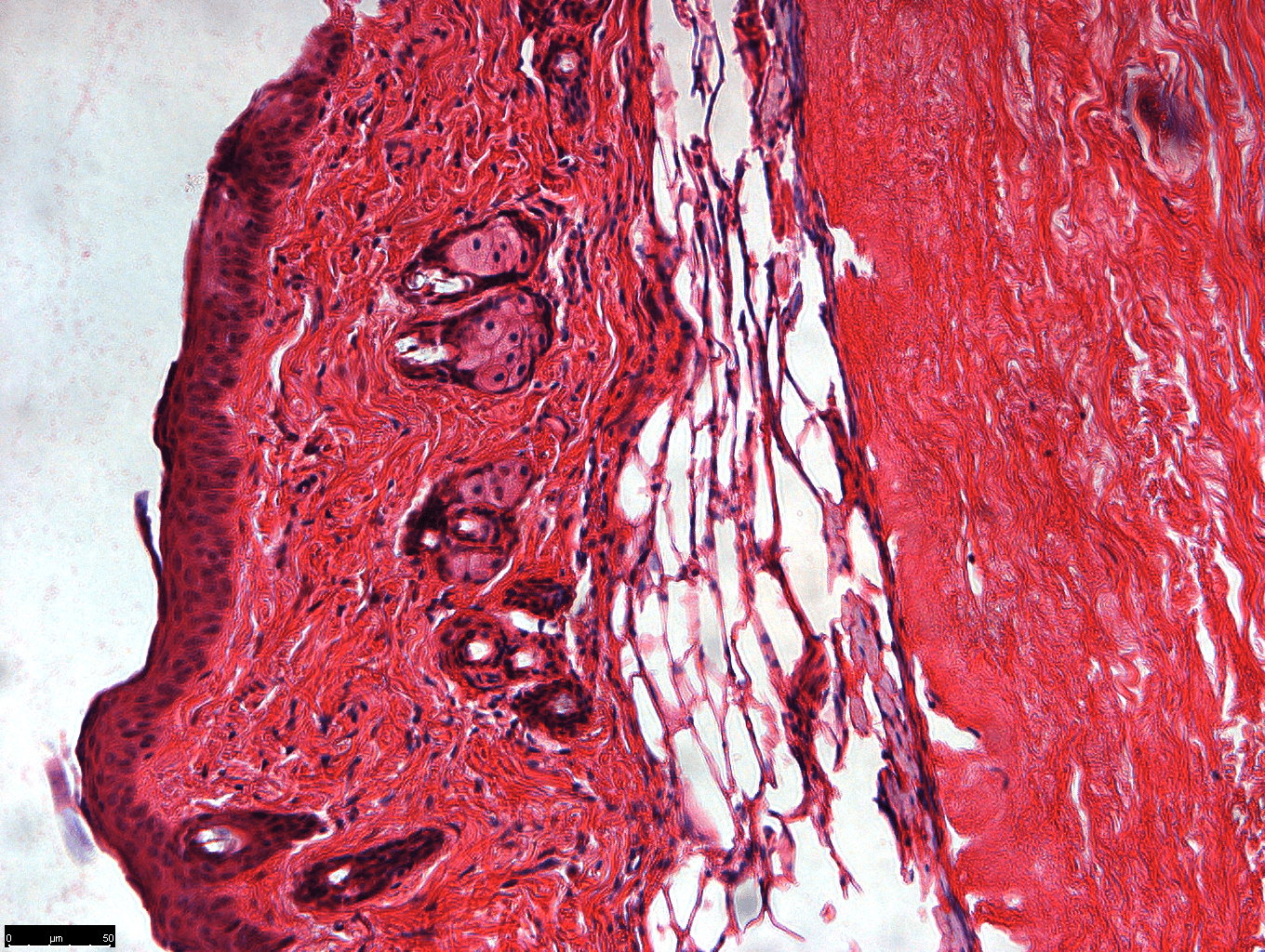
Mouse dermis, epidermis, and fat layers imaged with a more standard bright field microscope.
Green cells express GFP (green fluorescent protein).
